Diagnostics, including biopsy and Digital imaging
Biopsy
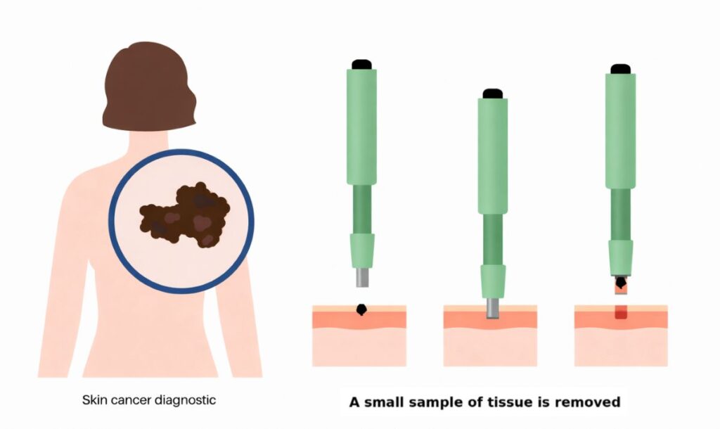
If any suspicious lesion is identified on clinical examination or dermoscopically, then a biopsy may be performed to take a sample (biopsy) for further analysis of the lesion at the histopathology lab.
This may include:
- Punch biopsy – taking a circular piece from the lesion of concern, usually around 3-4 mm in diameter
- Shave biopsy – this includes shaving off the lesion superficially, using a surgical blade, can be used for clinically superficial lesions
- Excision biopsy – sometimes it is ideal to remove the entire lesion for histopathological examination, especially for moles with concerning features.
Biopsy provides useful information regarding the type of skin cancer, the depth of skin cancer and also examines any risk features which allows planning of treatment options and clinical margins if surgery is required.
Digital imaging (photography) of skin lesions
Digital imaging (photography using computers) is an important aid in the monitoring, evaluation and detection of skin cancers. We use digital monitoring to:
- Closely examine skin lesions at high (30x to 50x) magnification
- Monitor skin lesions over time, to detect suspicious changes
- Automatically analyse characteristics of skin lesions such as symmetry, borders, size, presence of suspicious colours and structures
- Record the location of skin lesions in selected body areas (or the whole body) and pinpoint new or changing skin lesions
Based on the photographs of your skin lesions, we can decide whether:
- There is a reasonable possibility of skin cancer and the spot must be removed
- The chance of skin cancer is low, and the spot may be kept under observation and re-photographed later.
We use the latest imaging technology which allows us to easily observe the characteristics of your skin lesion and check for changes in your spots over time.
Procedures such as biopsies, excisions and cryotherapy are charged separately and quoted at your consultation. Medicare rebates apply where eligible. View full fees.